International Journal of Scientific & Engineering Research, Volume 3, Issue 7, July-2012 1
ISSN 2229-5518
HOW INSULIN REGULATES INSECT GROWTH: THE EVIDENCE
Dr. Barry.G. Loughton, Mr. Janpreet Singh Atwal
(Department of Biology, York University, 4700 Keele Street, Toronto, M3J 1P3 )
Abstract— Insulin is now recognized to be an important hormone of the invertebrate system. Insulin; after its discovery by Banting and Best in 1921 is now known to be present in almost all invertebrate species. It was first discovered in the pancreatic Islets of Langerhans of Canis familiaris. Insulin like activity was first documented in the bivalve mollusc Mmya arenaria. Insulin like peptides were purified from Bombyx heads Bombyxin II was the first molecule to have its amino acids sequenced. Bombyxin II showed significant similarity to the mammalian insulin and thus, was declared as insulin like molecule in invertebrates.
Index Terms— Bombyxin, Insulin, Insect insulin, Insulin like molecules
—————————— ——————————
INTRODUCTION
Insulin is now recognized to be an important hormone of the invertebrate system. Insulin after its discovery by Banting and Best in 1921 (Dominguez, 2001) is now known to be present in almost all invertebrate species. It was first discovered in the pancreatic Islets of Langerhans of Canis familiaris. Insulin like activity was first documented in the bivalve mollusc Mmya arenaria (Collip, 1923). Insulin like peptides were purified from Bombyx heads (Nagasawa, 1984). Bombyxin II was the first molecule to have its amino acids sequenced. Bombyxin II showed significant similarity to the mammalian insulin and thus, was declared as insulin like molecule in invertebrates.
THE INSULIN MOLECULE AND RECEPTOR
Mammalian insulin was discovered in 1921 and bovine insulin was sequenced in 1953. It was the first protein to be sequenced (Stretton, 2002). The insulin molecule consists of the pre, A, B chain and the C chains (Meyts et al, 2007). These four domains exist as pro insulin peptides without any biological activity. The pre and C domains get cleaved and pro insulin in mod- ified to produce biologically active insulin. The molecular weight of insulin is 6KDa which is relatively small for a poly- peptide. The A and the B chains are 21 and 30 amino acids long respectively (Permutt et al, 1981).The A and the B chains have receptor binding activity for the insulin substrate. The A and the B chain are joined together by two disulfide bonds contributed by cysteine residues. There is one disulfide bond within the A chain which is called the intra A chain disulfide bond. The bond forms early in the folding of the insulin mole- cule (Yuan et al, 1999) where it stabilizes the A chain. The dis- ulfide bonds between the A and the B chains cause the A and B chains to acquire the helical quaternary structure. The prop- erly folded quaternary structure has biological activity. All eukaryotes possess the basic folding pattern in the insulin and insulin like growth factors (IGFs) molecules. Ten members of insulin family are known to be found in humans (Meyts et al,
2007) like growth factors (IGFs) molecules. Ten members of
insulin family are known to be found in humans (Meyts et al,
2007).
Tyrosine kinases have modular domains with subunits that
are made in the cytoplasm by different genes and put together
and transported to the cell membrane through vesicles. The
insulin receptor exists as a dimer of two different types of chains namely the alpha and the beta chain.

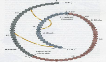
Fig 1: The primary structure of the pro insulin (Voet and Voet, 1995) molecule showing A, B and C chain. The two inter-chain disulfide bonds between A and B chains are shown. Cysteine 7 of B chain makes a disulfide bond with cysteine 7 of the A chain. Cysteine 19 of B chain makes a disulfide bond with cysteine 20 of the A chain. An intra- chain disulfide bond is present between cysteine 6 and cysteine 11 of the A chain. Proteolytic cleavage between glycine 1 of A chain and arginine 63 of C chain and proteo- lytic cleavage between alanine 30 of B chain and arginine
31 of C chain yields two chains that produce the active form of insulin. The inter and intra disulfide bonds help in correct folding of the protein as well as imparting biologi- cal activity to the insulin molecule.
The expression and identification of the insulin receptor was first documented in guinea pigs and humans (Zhang et al,
1992). There are 22 exons and 21 introns in the mammalian insulin receptor gene. Exons are sequences of nucleotides that are made into mRNA by RNA polymerases. Introns are nuc- leotide sequences that are not expressed and get spliced out during post translational modifications through a process called alternative splicing. The insulin gene gives rise to the alpha and the beta chains. The addition of carbohydrate oli- gomers to the alpha and the beta chains occurs in the cytosol. After glycosylation, the alpha and the beta chains are trans- ported to the Golgi apparatus by calcium binding proteins
IJSER © 2012
http://www.ijser.org
The research paper published by IJSER journal is about HOW INSULIN REGULATES INSECT GROWTH: THE EVIDENCE 2
ISSN 2229-5518
calnexin and calcineurin. In the Golgi apparatus, the glycosy- lated chains are dimerized to form the mature α2β2 tetramer which is transported to the plasma membrane of the cell. The insulin molecule has a very complex pathway downstream of the insulin receptor. Insulin molecule can modulate many functions like cell growth, glucose metabolism and immune response through the modular structure of insulin receptor.
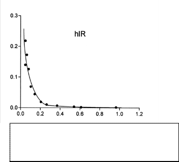
Insulin has a high affinity to its receptor. The membrane bound insulin receptor has a dissociation constant Kd of
6.3x10^-9 M.
ECM
Plasma membrane
Fig 3: Scat chard plot (Peter et al, 2000) showing the binding affinity assay for insulin receptor. The dissocia- tion constant Kd for the insulin receptor was found to be 6.3x10^-9 M which is indicative of very high affinity.
Trans- membrane domain
Cytoplasm
MAMMALIAN INSULIN
The most crucial role of insulin in mammals appears to be the regulation of glucose metabolism due to strong correlation between insulin and occurrence of diabetes (Perkins and Rid- dell, 2006). The insulin molecule binds to its receptor which causes the cytoplasmic domains of the receptor to get auto- phosphorylated. This autophosphorylation further cross phosphorylates the adjacent tyrosine residues which now can recruit other proteins. The phosphorylated proteins then bind Phospho-inositide 3 Kinase which binds to the Grb2 and mSOS protein on the phosphorylated tyrosine sites. The mSOS and Grb2 complex further binds SHP-2 protein which is a ty- rosine phosphatase. The function of SHP2 is to dephosphory- late the tyrosine residues to shut off the signal transduction
Fig 2: The insulin receptor (Jongsoon and Pilch, 1994) has two chains with subunits namely the alpha and the beta subunits. The alpha subunit lies on the extra cellu- lar side .The alpha subunit has cysteine rich domains which help bind the insulin substrate. The dotted lines between the alpha and beta subunits show the disulfide bonds. There is a trans-membrane domain in the beta subunit that anchors the insulin receptor in the plasma membrane. The trans-membrane region is rich in hy- drophobic amino acids which allow tight interaction of the receptor with the hydrophobic region of the cell membrane hydrophobic region. There is an ATP bind- ing region at lysine 1018 of the beta chain. The juxta membrane region has two phosphotyrosine sites whe- reas there are three phosphotyrosine binding domains
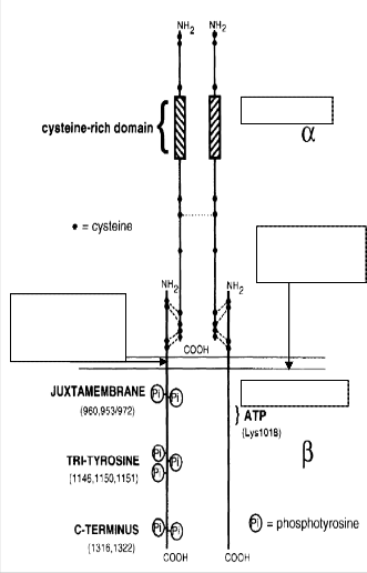
once the receptor has been activated. The activated tyrosines phosphorylate the membrane bound Phospholipase C to acti- vate PIP2 which gets cleaved into Diacyl Glycerol (DAG) and Inositide Phosphate 3 (IP3). Downstream of IP3: Protein Ki- nase B (PKB) activates Glycogen Synthase Kinase 3 beta through phosphorylation. Insulin also increases the intracellu- lar concentrations of cAMP by inhibiting cAMP phosphodies- terase. cAMP is the primary 2nd messenger downstream of the RTK receptor domains. GSK3beta facilitates the conver- sion of glucose into glycogen and also increases the uptake of glucose by transporting GLUT 4 receptor to the surface of the cell. Phosphorylation of GSK3beta reduces its kinase activity and thus increases its enzymatic activity to convert glucose in glycogen. The Grb and mSOS complex is involved in activat- ing the Ras-MAPK pathway which is involved in cell prolife- ration and growth. In this way, insulin maintains the glucose

IJSER © 2012
http://www.ijser.org
The research paper published by IJSER journal is about HOW INSULIN REGULATES INSECT GROWTH: THE EVIDENCE 3
ISSN 2229-5518
balance in blood by activating glycogen synthase kinase 3 be- ta. This pathway in mammals suggests that analogous molecu- lar mechanisms to control blood sugar levels perhaps be present in insects as well.

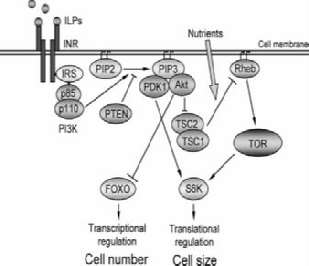
Fig 4: The diagram (Wu and Brown, 2006) shows the basic mechanism through which the insulin substrate elicits an intracellular response. The scaffolding and adaptor proteins such as p110 and p85 affect Phospha- tidylinositol (3, 4,5)-trisphosphate (PIP3) which through the Akt/ PDK regulate cell size and growth. The p85 and p110 adapter proteins bind to the phosphotyrosine domains of the cytoplasmic domains of the beta subunit of the insulin receptor. Insulin is involved in glucose metabolism, cell survival, and cell division.
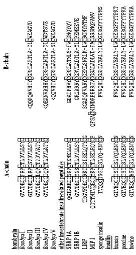
tween the amino acid sequences of insulin alpha and beta chains from various invertebrates including sponges with the mammalian insulin alpha and the beta chains. The highly con- served nature of the cysteine residues indicate the importance of the disulfide inter bridge that is used to hold the insulin alpha and beta chains together.
CONSERVED NATURE OF INSULIN MOLECULE
Many phylogenetic and molecular studies show that almost all species have insulin in them. Sequencing and structure analy- sis (Nagata et al, 1995) showed significant similarities in the Bombyxin A and B chains compared to human (homo sa- piens), mouse (Mus musculus), fruitfly (Drosophila melanogaster) and Nematode (C.elegans). For the insulin protein quaternary structure, all species studied show the basic proline rich fold and two inter-chain disulfide bonds which are hallmarks of the insulin molecule. The cysteine residues are known to be highly conserved in the molecules of insulin like molecules which include insulin, insulin like growth factors (IGFs) and relaxins. Nagata et al, 1995 researched the relationship be-
Fig 5: The above figure (Nagata et al, 1995) shows the sequence of the alpha and beta insulin chains in the silkmoth (Bombyx mori), sponge (Geodia cydo- nium), mollusc (Lymnaea stagnalis), human (homo sapiens), pig (Sus scrofa), bull (Bos taurus), locust (Locusta migratoria). The Bombyx I, II, II, IV, V are insulin related peptides from the arthropod Bombyx mori. Insects have multiple genes for insulin while mammals possess only one type. SBRP1A, SBRP1B are the insulin related peptides from Samia (Samia cynthia). LIRP is the locust insulin related peptide. MIP I is the insulin mollusc insulin related peptide I. It can be clearly seen that all the cysteines in the A and the B chains are conserved. The cysteine resi- dues are important in connecting the A and B chains of insulin related peptides together.

IJSER © 2012
http://www.ijser.org
The research paper published by IJSER journal is about HOW INSULIN REGULATES INSECT GROWTH: THE EVIDENCE 4
ISSN 2229-5518
halose and glucose levels in the haemolymph of Calliphora
vomitera.
Nematode
Interchain disulfide bonds
Glial cells
Neuropile
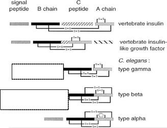
Fig 6: Duret et al, 1998 compare the A, B and C chains of the insulin molecule in vertebrates and in nematode C.elegans which is a primitive organism. The diagram shows that the disulfide inter-bridge is conserved in C.elegans insulin as well in verte- brate insulin.
Insulin like reactivity with bovine antiserum

IMMUNOCYTOLOGICAL EVIDENCE OF INSULIN IN INSECTS
Initially, insulin was looked upon only as a metabolic hor- mone. Control of sugar metabolism was the main function that was attributed to the insulin peptide. Insulin isoforms such as insulin growth factor 1 and insulin growth factor 2 (IGF1 and IGF2) have been shown to promote growth and proliferation of cells by affecting the Akt/PDK and TOR pathway (Wu and Brown, 2006). Insulin itself is now also considered as an ana- bolic hormone due to its facilitation of the insect growth.
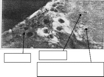
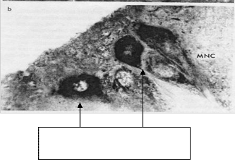
In insects, insulin was observed to regulate trehalose levels in the haemolymph of Calliphora. Duve and Thorpe, 1979 were able to localize insulin like material in the Median Neurosecre- tory cells of the blowfly, Calliphora vomitoria. The MNC is lo- cated in the protocerebrum of the insect brain. Through im- munocytological staining, the researchers were able to visual- ize an insulin like substance that reacted with bovine insulin antibodies that were derived from guinea pigs. The research- ers ablated the Median Neurosecretory cells from the insect Calliphora vomitera. As a result, the insect became hypertreha- losemic and hyperglucacemic half an hour after the removal of the neurosecretory cells. However, when the insect was in- jected with bovine insulin, the trehalose and glucose levels had returned to normal. These findings led the researchers to establish that insulin like peptides were localized in the me- dian neurosecretory cells and were involved in lowering tre-

Insulin like reactivity with bovine antiserum
Fig 7: The above pictures (Duve and Thorpe, 1979) show the pars intercerebralis of the protocerebrum of Callipho- ra vomitoria. The first picture shows the regions where the MNC cells, glial cells and the neuropile are located. The second picture shows the darkened regions near the neuropile and MNC. These stains were due to the im- muno-reactivity of bovine insulin antiserum with the insulin like material present in the darkened region.
INSULIN LIKE PEPTIDES AND MOULTING
Sevala et al, 1991 showed the presence of insulin like material in Rhodnius prolixus by using anti-bovine insulin serum pro- duced in guinea pigs. The researchers prevented moulting in the 5th stage of Rhodnius larvae by injecting anti-bovine insu- lin serum. The researchers injected insulin immediately after the larvae had fed and on days 4 and 5 in females. The re-
IJSER © 2012
http://www.ijser.org
The research paper published by IJSER journal is about HOW INSULIN REGULATES INSECT GROWTH: THE EVIDENCE 5
ISSN 2229-5518
searchers injected the insulin antiserum in male larvae on days
5 and 6. The result was that all these larvae had failed to
moult. The researchers used control antiserum from rabbits
which were challenged with crustacean red pigment. Those
insects which received the control antiserum were able to
moult successfully. Thus, they could prove the existence of a
substance that reacted with insulin antiserum.
Protoce- rebrum on day-2
Day-
3protocerebrum Protocerebrum of
day-5 pupa
Corpora allata of day-0 pupa
Protocerebrum of day-1 pupa
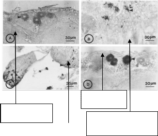
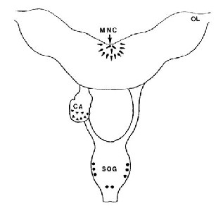
Fig 8: The picture on the left (Sevala et al, 1992) shows the general structure of insect brain. Optic lobes are labelled as OL. MNC is the Median Neurosecretory Cell region. CA is the Coprus Allatum. SOG is the Subesophageal Ganglion. The areas with dark dots and triangles show regions of immunoreactivity with insulin antiserum. MNC, CA and SOG showed insulin specific immuno- reactivity.

Day-3 pro- tocerebrum
Late larva pro-
tocerebrum Protocerebrum of day-0 pupa
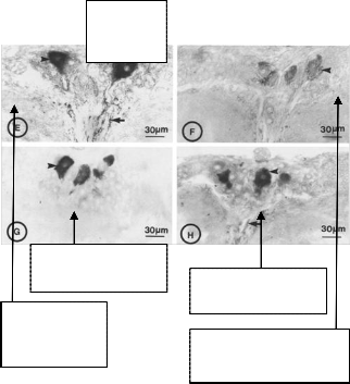
Fig 9: The picture above (Sevala et al, 1992) shows the variance in intensity of insulin specific immunocyto- logical staining on different days of pupal develop- ment in beetle. The staining was done in the neurose- cretory cells and the axon termini of in the protocere- brum and retrocerebral complex of Tenebrio molitor. The arrows above indicate the axon termini and the arrowheads alone in the pictures indicate neurosecre- tory cells.

INSULIN LIKE PEPTIDES AND MOULTING
Sevala et al, 1991 showed the presence of insulin like mate- rial in Rhodnius prolixus by using anti-bovine insulin serum produced in guinea pigs. The researchers prevented moulting in the 5th stage of Rhodnius larvae by injecting anti-bovine in- sulin serum. The researchers injected insulin immediately after the larvae had fed and on days 4 and 5 in females. The re- searchers injected the insulin antiserum in male larvae on days
5 and 6. The result was that all these larvae had failed to moult. The researchers used control antiserum from rabbits which were challenged with crustacean red pigment. Those insects which received the control antiserum were able to moult successfully. Thus, they could prove the existence of a substance that reacted with insulin antiserum.
In Tenebrio molitor (Sevala et al, 1992) the insulin like pep- tides are released from parts of the protocerebrum and retro- cerebral complex and chiefly in the Median Neurosecretory cells. Insulin like material is also produced in the corpus alla- tum. Remarkable changes were observed in the concentration of the insulin like peptides in the Median Neurosecretory Cells during the early stages of the pupal development of the beetle.
IJSER © 2012
http://www.ijser.org
The research paper published by IJSER journal is about HOW INSULIN REGULATES INSECT GROWTH: THE EVIDENCE 6
ISSN 2229-5518
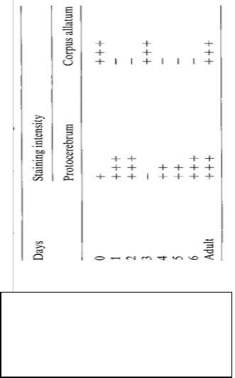
The protocerebrum showed weak staining on day 0 of the pupa. However, on the second and the third days, the proto- cerebrum showed strong immunological reactivity to anti in- sulin antiserum. Almost, negligible staining was observed on the third day whereas the staining intensity increased on the fourth day until day six which showed strong immunoreactiv- ity resulting in dark stains. In corpus allatum on the other hand, the staining pattern was different than that of the proto- cerebrum. Day zero showed strong staining patterns. There was weak staining in the corpus allatum on the second and the third days. Again on the fourth day, strong staining was shown followed by no staining on the fourth, fifth and the sixth days. The adult corpus allatum however showed strong staining which was similar to that on day zero.
Fig 10: Anti insulin specific immunoreactivity (Sevala et al, 1992) in the protocerebrum and corpus allatum of the beetle Tenebrio.The immunocytological staining was performed from day zero to adulthood in the beetle. – shows no immunoreactivity, +++ show strong immunoreactivity and ++ show moderate im- munoreactivity.
The researchers were able to delay the pupal development by injecting the insulin antiserum in the protocerebrum of the beetle. The studies showed that the insects were the most sen- sitive to insulin antiserum immediately after larval and pupal ecdysis. However, after five hours of ecdysis, the anti insulin serum had no effect on the duration of the instar. The anti in-
sulin antiserum when injected on the second and the third day caused the insects to have longer stadium especially on the third day. Such evidence led these researchers to believe that insulin like substance might have played a role in regulating the length of the instar and hence insect growth.
Older studies on insect moulting have suggested the role of nutrition and hormones in the moulting cycle. A study (Beckel and Friend, 1964) showed that stomach distension due to feed- ing in Rhodnius could initiate moulting. They conducted expe- riments on the Rhodnius by feeding the insects different types of meals consisting of saline solution and saline with dextran. They found that both of these solutions caused the insect to undergo moulting since these solutions were able to stretch the walls of the insect‘s stomach. The stretching of abdominal muscles in the insects caused the initiation of mitosis in epi- dermis. However, there were no direct conclusions about presence of insulin in insects and its role in insect growth.
Interesting findings about the role of nutrition in develop- ment have been well recorded in the insect Rhodnius prolixus. Histological studies (Mulye and Davey, 1995) have showed the presence of a humoral factor in the haemolymph of the insect which was transmitted to the neuroendocrine axis in the brain that caused the cells to increase their action potential frequency. The researchers showed that, on severing the dor- sal vessel in the insect circulatory system, action potential fre- quency in the cells that makeup the neuroendocrine axis was significantly reduced. The dorsal vessel is the most important in hemolymph circulation since the direction of the flow is from the abdomen to the brain. The dorsal vessel carries the majority of the hemolymph from abdomen to the brain of the insect. The researchers also noted that severing the dorsal ves- sel greatly reduced the number of eggs produced compared to insects which had an intact but exposed dorsal vessel. The experiment provided direct evidence for the hypothesized humoral factor that carried the feeding signal to the brain. Ex- tensive studies on Rhodnius physiology (Wigglesworth, 1934) showed that severing of the dorsal vessel of the fifth stage lar- vae delayed moulting. However, Orchard and Steel, 1980 measured the effect of severing the dorsal vessel on the action potential frequency in corpus cardiacum. Through their stu- dies on action potentials in corpus cardium, Orchard and Steel, 1980 had already established that increase in action po- tential frequency was as an index of the prothoracicotropic hormone release. The researchers suggested that the signal to release prothoracicotropic hormone was also through the hu- moral route. Mulye and Davey, 1995 tried to clarify these con- ceptions through their unpublished data. They showed that diuretic hormone release controlling diuresis was unaffected by severing the dorsal vessel in Rhodnius. From such evidence, they suggested the presence of two different mechanisms re- sponsible for development and maintenance of homeostasis such as through diuresis after consumption of a large blood meal by Rhodnius.
DROSOPHILA INSULIN LIKE PEPTIDES (DILPS)
IJSER © 2012
http://www.ijser.org
The research paper published by IJSER journal is about HOW INSULIN REGULATES INSECT GROWTH: THE EVIDENCE 7
ISSN 2229-5518
Modern studies have followed the evidence on insulin like immunoreactivity in the protocerebrum in insects. Recent re- search has been focused on the Drosophila insulin receptor (Dinr) and its function in insect fat bodies and in controlling hemolymph sugar levels (Okamoto et al, 2009). Although, it had been already known that insulin like peptides can affect sugar levels in invertebrates yet other questions remained. Research (Okamoto et al, 2009) had shown that insulin like peptides could regulate growth in insects. The researchers examined various Drosophila insulin like peptides namely, dilp
1, dilp 2, dilp 3 dilp4, dilp5, dilp6 and dilp7. Drosophila insulin like peptides 1, 2, 3 and 5 were found in the brain neurosecre- tory cells of the fruitfly. Dilp 6 was found to be abundant in the fat body of the insect. Dilp 7 was found in the neurosecre- tory cells of the ventral nerve cord (Nijhout and Grunert,
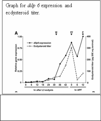
2002). The researchers examined the fat body of the Drosophila pupa till reaching the adult stage for dilp expression by using RT-PCR (Real time polymerase chain reaction) to quantify the expression of the dilp genes. The researchers found high levels of dilp 6 in the fat body of the pupa less than one hour after pupa formation. On studying the levels of ecdysteroid in the late third instar of Drosophila larva, dilp6 expression was paral- lel to the levels of 20-hydroxyecdysone. It was found that the
20-hydroxyecdysone directly influenced the dilp 6 levels. In order to confirm the relationship between dilp6 levels and 20E the researchers added cycloheximide, a protein synthesis inhi- bitor in vivo. The addition of cycloheximide eliminated the effects of 20E induced transcription factors. However, the ex- pression of dilp 6 was still induced in the presence of cyclohex- imide. The dilp6 levels continued to rise with the rise on 20E levels. 20E is a key hormone related to moulting in insects. This showed that dilp 6 could directly initiate 20E production.
DILPS AND INSECT GROWTH
Also, in Drosophila, insulin has been suggested to play a role in the proliferation of and development of imaginal discs in the pupal insect (Bryant, 2008). Imaginal discs are structures that are present in the pupa that grow rapidly during metamor- phosis. The imaginal discs give rise to wings, legs and anten- nae during metamorphosis. Bryant, 2008 demonstrated that the insulin receptor stimulation was responsible for the rapid growth of imaginal discs in Drosophila. The study showed that the IDGFs (Imaginal Disc Growth Factors) from the fat body activate the insulin receptor pathway by stimulating the insu- lin receptor. The IGDFs are glycoproteins that modulate vari- ous types of signalling cascades and produce pro inflammato- ry responses that can activate the insulin receptor. Based on nutritional availability, the insulin/IGF had a general effect on cell proliferation and cell growth when IDGF signal was also present. IGDFs are related to chitinase enzymes that break down chitin but they can undergo an amino acid substitution to nullify the chitinase activity and convert into lectins. These endogenous lectins then exert their nutrition based effects on the growth through the insulin receptor pathway.
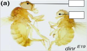
Intensive studies in Drosophila have shown that insulin regu- lates growth in Drosophila. Brogiolo et al, 2001 were able to mutate Drosophila insulin homologue Dinr gene. Dinr controls the cell number and size in a cell-autonomous manner. The researchers induced a partial loss of function mutation in the Dinr gene. As a result, the adult insects were much smaller in size than flies that had the normal Dinr gene. They found that the phenotype of the mutant flies was similar to the flies which had an impaired PI3K/PKB pathway. Thus, the re- searchers could conclude that Dinr regulates growth atleast at the PI3K/PKB level. On the contrary, the researchers were able to induce dilp2 overexpression by using hs-Gal4 driver line. It was observed that the flies with the overactive dilp2 gene were larger in size than normal flies. The flies with in- creased dilp2 were 39% heavier than the normal flies. The flies also had 5% more ommatida and had a wing area that was
21% larger than that of normal flies.
Normal
Mutant
Fig 11: Okamoto et al, 2009. Graph shows a close relation between the levels of ecdysteroid and dilp6 in fat body of Drosophila larva. The data was taken within the first hour ecdysis after the third instar of larva. The levels of dilp6 and ecdysteroid are almost parallel.
SER © 2012
http://www.ijser.org
The research paper published by IJSER journal is about HOW INSULIN REGULATES INSECT GROWTH: THE EVIDENCE 8
ISSN 2229-5518
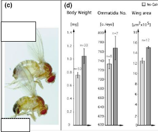
Fig 12: The bigger fly on the left was used as con- trol and had a normal working Dinr gene. The adult achieved a normal body size. The smaller fly on the right had a mutated Dinr gene with partial loss of function. The adult mutant fly was signifi- cantly smaller than the flies with a normal Dinr gene.

High
dilp2

Normal
Fig 13: The top fly on the left has a bigger body size, more wing area and larger number of omma- tidia. The fly below the mutant is a fly with normal dilp2 expression. The mutant was 39% heavier and had 5% more ommatidia. The wing area increased by 21% compared to normal wing size. The bar diagrams show comparison between the average values of body weight, ommatidia number and wing area between the normal and mutant dilp2 flies.
Another later study (Tu et al, 2005) provided very convincing data about the role of insulin in longevity and overall adult size in Drosophila. Drosophila heteroallelic mutants with mutant Dinr were infertile, smaller in size than normal adults and also lived longer than normal wild type Dinr containing flies. Also, the researchers produced homozygotes with genotype InRE19/InRE19 which had very low levels of Juvenile hor- mone. Also, the researchers produced flies that were mutated homozygotes with the genotype chico1/chico1. The chico pro- tein acts as a substrate for the Drosophila insulin receptor. By acting downstream of the Drosophila insulin receptor, chico stimulates the insulin pathway and leads to cell growth and proliferation. Dinr mutant flies had smaller than normal size and showed similar physiological states when compared to the InRE19/InRE19 and InR p5545/InRE19. The researchers
were able to rescue the mutant flies by application of Juvenile hormone analogues. The rescued Dinr mutant genotypes were able to show normal wild type lifespan and vitellogenesis. According to the researchers, the insulin signalling may con- trol the neurons in the brain that control the synthesis of Juve- nile hormone. The researchers also found that the regulation of JH is influenced by insulin dependent PI-3 kinase pathway. Oldham et al, 2002 had earlier proved in 2002 that chico and Inr function through the PI3 Kinase pathway to induce cell growth and proliferation.


Fig 14: Molecular pathway (Garofalo, 2002) showing the action of Drosophila insulin like peptides on the insulin receptor to cause cellular growth.
Bombyxin (Nijhout and Grunert, 2002) was extracted from the silk moth Bombyx mori and its function on growth was studied. The researchers used the wing imaginal discs from the butterf- ly Precis coenia as a growth model. The wing imaginal discs were removed from the larva and kept in vitro in a culture medium. Haemolymph from the extract from Precis coenia and Manduca sexta and active bombyxin in the haemolymph was collected. 20-hydroxyecdysone was also present in the haemo- lymph extract but was not sufficient enough to support moult- ing. The larvae from Precis coenia were at the fifth instar of their life cycle when the imaginal discs were taken from them and fixed in vitro. At the fifth instar in Precis coenia, the im- aginal disc cells of the wings divide at an exponential rate. The researchers used guinea pig and mouse monoclonal antibodies to Bombyxin in the haemolymph that supported the imaginal disc growth in vitro. The researches then used the haemo- lymph containing the monoclonal antibodies over an antibody affinity column to separate the bombyxin-antibody complex from the haemolymph. Then they used the same eluted hae- molymph to support the imaginal disc growth. As predicted, the researchers found that the haemolymph without bombyx- in was unable to elicit any growth response in the wing im-
IJSER © 2012
http://www.ijser.org
The research paper published by IJSER journal is about HOW INSULIN REGULATES INSECT GROWTH: THE EVIDENCE 9
ISSN 2229-5518
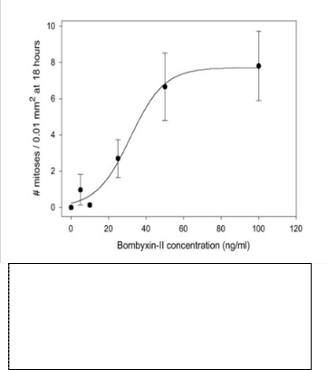
aginal discs. Following their observations, the researchers decided to add 50 ng/ml of artificially made Bombyxin II. Bombyxin II is an isoform of Bombyxin found in the silk moth Bombyx mori. The addition of the Bombyxin was able to restore growth in the wing imaginal discs of Precis coenia in the pres- ence of 20E. Both Drosophila and Precis require the involve- ment of insulin (bombyxin) in the wing imaginal discs for their growth and proliferation.
Fig 15: Nijhout and Grunert, 2002 shows the number of cell divisions in the imaginal disc cells over different concentrations of bombyxin II in the haemolymph of Precis coenia. The number of cell divisions increases al- most exponentially from 0-60ng/ml of bombyxin II. The number of cell divisions did not increase from con- centrations from 60 ng/ml to 100ng/ml.
MOLECULAR BASIS OF DILP ACTION
Cells grow in volume and surface area before they divide. Once a cell reaches a certain volume to surface ratio, it com- mits itself to divide. All cells need nutrients that are supplied through the diet of the animal. Much evidence shows that in- sulin production is directly related to nutrient availability (Oldham et al, 2002). The plot showing the number of cell di- visions versus bombyxin II concentration reached a saturation point after 60ng/ml. Researchers (Kaplan et al, 2008) found a GTPase called ns3 which strongly regulates the insulin path- way and ultimately the body size in Drosophila. GTPases are proteins that deliver energy for intracellular signalling cas- cades and hydrolyze GTP to GDP and inorganic phosphate. The GTPase ns3 or nucleostemin 3 is a GTPase present in the nucleolus of the Drosophila embryonic cells. The researchers used a Drosophila strain which had a P element in the ns3 gene. The insertion of the P element led to the loss of function of the gene. The mutant created, lacked a functional ns3 gene. The second exon of the ns3 gene contained the P element. The
studies showed that the ns3 mutants did not affect embryonic development but severely retarded the larval growth. The adult ns3 mutants had normal lifespan but had only 60% of the body weight relative to the adult wild type flies with a functional ns3 gene. The loss of ns3 function also retarded the growth of imaginal discs and led to the formation of smaller wings on the adult mutants. These results were confirmed by a rescue experiment further. The researchers introduced a functional ns3 gene leading to normal development of the mu- tant flies when compared to the normal flies. They also found that the Akt kinase activity which lies near the end of the insu- lin signalling cascade was deregulated in the ns3 mutant lar- vae. Akt is phosphorylated on receiving a stimulatory signal from insulin. However, in the case of ns3 mutants, Akt could not be phosphorylated which reduced ribosome de novo syn- thesis. The researchers found low levels of ribosomes in the ns3 mutants compared to the normal flies. The peripheral in- sulin pathway was inhibited which led to growth retardation in the ns3 mutants. The researchers linked the growth defects in the ns3 mutants to the impairment of serotonergic neurons in the Drosophila central nervous system which are linked to MNC which secrete Drosophila insulin like peptides. The re- searchers were able to rescue the ns3 mutants to normal size and lifespan by implanting serotonergic neurons that restored the normal phosphorylation of Akt kinase downstream of the insulin pathway. Nutrition is a key limiting factor in the in- sects that controls the insect‘s growth. Studies in Drosophila have provided robust evidence that nutrition controls the growth and determines adult size. The fat body of Drosophila is responsible for the production of insulin like growth factor dilp6. Okamoto et al, 2009 also showed how dilp6 peaked in the fat body of Drosophila immediately after feeding.
The prothoracic gland is an important endocrine hormone in insects which plays a key role in the onset of moulting. Studies done is Drosophila have provided key insights how the Protho- racic gland acts as a sensor of the insect‘s body weight and nutrient availability. The insect has to reach a certain weight called the critical weight before it begins the moulting process. Reaching the critical weight is essential since it is reflective of the nutrients required to support metamorphosis. This me- chanism is an ―all or none‖ phenomenon. Studies in deter- mining the critical weight of insect prior to moulting have been done in the tobacco hornworn (Manduca sexta) and Dro- sophila melanogaster. In 1975, Nijhout examined metamorphosis in Manduca sexta to understand the underlying mechanism that committed the insect to undergo metamorphosis. Nijhout,
1975 described that in Manduca, metamorphosis did not occur
until the insect attained a species specific size. The researcher
used 5th instar larva since most noticeable growth occurred in this stage. Nijhout, 1975 measured the size of the head cap- sules of the insects to record growth in the insect. The re- searcher starved the Manduca larva when these weighed un- der 3 g. Only a few larvae were able to proceed to the next instar. However, these larvae were unable to form pupae. The researcher also starved another set of Manduca larvae which weighed under 4 g. Many of these insects were able to molt but transformed into non-viable larval-pupal intermediates.
IJSER © 2012
http://www.ijser.org
The research paper published by IJSER journal is about HOW INSULIN REGULATES INSECT GROWTH: THE EVIDENCE 10
ISSN 2229-5518
When larvae that weight over 4 g were starved, many of these insects were able to transform into pupae but the pupae were When 3rd and 4th instar larvae were temporarily starved and allowed to moult at a subnormal weight, the resulting 5th in- star larvae were proportionally smaller. Only larvae which had head capsules that were 5.1 mm or higher were able to form normal size pupae and proceed to the next instar. The researcher found that the minimum size to form a normal pu- pa was 5.1 mm for the head capsule of the insect. Thus, the researcher was able to establish an accurate relationship be- tween size and weight of insect that acted as the threshold point for moulting initiation. (Mirth et al, 2005) argued that the determination of critical weight was done through the Drosophila insulin receptor by chico substrate protein for the Dinr that precedes PI3 (Dp110) kinase activation through p60 its adapter protein. The critical weights in Drosophila could be divided into pre and post critical weights. The Drosophila insu- lin receptor plays different roles in the pre critical weight and the post critical weight periods. In the pre critical weight pe- riods, the Dinr influences the time of larval development and does not influence body size. In the post critical weight pe- riods, Dinr does not influence the time of larval development but regulates body size. These functions of Dinr were nutrition based according to the researchers. Mirth et al, 2005 were able to show a correlation between the size of prothoracic gland and critical weight that would be required for insect to initiate metamorphosis.
The researchers were able to produce prothoracic glands in Drosophila which were precociously enlarged by using phan- tom based increased Dp110 expression. The results of such modifications were convincing. The P0206 GAL4 to drive UAS PTEN induced early metamorphosis in more than 50% of the insects to progress to second instar (L2) pupa. But than 1% of the L2 pupa formed pharate adults which had formed a new exoskeleton at the end of second instar. Therefore, 34% of the larvae progressed to third instar (L3) to form pharate adults. The P0206 >UAS PTEN lines also showed developmental de- lays to reach the second instar (L2). Only 35% of the total indi- vidual larvae with the PTEN expression system were able to proceed to the L2 stage. In the control individuals, 90% of lar- vae proceeded to the L2 stage 2 days after hatching. More than
50% of the insects with suppressed PG showed delayed devel-
opment. The control larvae only took 24 hours in order to
progress to the third instar pupae whereas the PTEN lines took 3 days to progress to the third instar pupae.
In order to establish a link between nutrient intake and devel- opment, the researchers also fed the PTEN larvae food con- taining different nutrients. A greater number of larvae that
were underfed at the L2 stage were able to form the L3 pupae. However, phantom>Dp110 larvae with enlarged prothoracic glands showed no difference in size at the L1 and L2 stages. There was no difference between the duration of the L1 and L2 stages of these insects compared to the control individuals. The researchers determined the Minimum Viable Weight of phm>Dp110 L3 larvae. Minimum viable weight is the mini- mum weight attained by 50% of the larvae to survive and un- dergo metamorphosis. Starved phm >Dp110 larvae were una-
ble to form pupae. Only 50% of these individuals were able to form pupae after 11.5 hours with weight of 0.52mg. The Min- imum Viable Weight to initiate pupa formation of the control larvae was measured to be 0.88mg after a period of 11.5 hours. Since the researchers did not find any significant differences in the duration of the L1 and L2 stages in the phm>Dp110 lines when compared to the control individuals, they decided to measure ecdysteroid levels in the phm>Dp110 early L3 feed- ing stage. Surprisingly, they did not find any significant dif- ferences between the ecdysteroid titers of the phm>Dp110 and the control individuals. It was clear from the PG size variation experiment, that reduced size of the prothoracic gland in- creased the duration of each instar while producing larger adults than normal. The researchers showed that growth of phm>Dp110 was nutrition based and the size of prothoracic gland served as a sensor to assess nutrient levels in the insect that would allow progression to the next instar. All well fed larvae having the phm>Dp110 with enlarged prothoracic glands showed shorter instar periods than the PTEN sup- pressed larvae especially in the L3 stage. These well fed larvae showed the same growth rate compared to controls.
Insulin controls moulting in locusts
Unpublished data on locusts (locusta migratoria) suggests that there is a critical time during the feeding when moulting cycle is initiated. In the experiment, 5th instar locusts were main- tained on a light dark cycle of 12 hours. The temperature dur- ing the dark part of the cycle was kept at 22 degrees Celsius and the light temperature was kept at 36 degrees Celsius. The locusts were fed on wheat seedlings and wheat bran. The in- sects were starved on different days. The insects were starved on day 0, 1, 2, 3, 4, 5 and 6. On average, the insects in the 5th instar showed a duration of 8 days for when the insects were starved either on day 0, 1, 2, 4, 5 or 6. However, the insects that were starved on the third day and fed on days 0, 1, 2, 4, 5 and 6 showed instar duration of 9.7 days. This was significant compared to other instar duration periods. The data suggested a critical period on day 3 that failed to receive a feeding asso- ciated stimulus. In another set of 5th instar locusts, three groups of the insects were taken. The 1st control group insects were fed daily and its instar period was 7.6 days. The second control group that was fed all days except on day 3 had an instar duration of 9.6 days. The third group of insects was fed all days but not on the fourth day. The instar duration was 8.3 days for this group. The researches tried to rescue the insects that were starved on the 3rd day of feeding by injecting insu- lin. They used 100 ng, 300 ng, 500 ng, 1 ug, 300 ug A chain and
1ug B chain insulin in all the three groups. Insulin injected on
any other day than day 3 did not affect the duration of the 5th
instar. However, in the day 3 starved locusts, the researchers were able to shorten the instar duration of 9.6 days to approx- imately 8 days by injecting insulin. Thus, the researchers were able to show that insulin release on day 3 was the required stimulus that committed the insect to molt.
INSULIN AND EGG DIAPAUSE
Furthermore, insulin in Drosophila controls egg diapause. Insu-
IJSER © 2012
http://www.ijser.org
The research paper published by IJSER journal is about HOW INSULIN REGULATES INSECT GROWTH: THE EVIDENCE 11
ISSN 2229-5518
lin in C.elegans is also involved in control of diapause. Di- apause (Williams et al, 2006) is an adaptation in Drosophila and other related insects of the phylum to survive unfavourable conditions. In diapause, Drosophila undergoes a quiescent state of low metabolic activity and formation of pre vitellogenic oocytes. In nematodes like C.elegans, when living conditions are unfavourable, the nematode produces dauer larvae which have low metabolic activity and can store large amounts of fat. Stressful conditions like overcrowding, low food supply in- itiate the dauer larva formation in C.elegans. Researchers (Gerisch and Antebi, 2004) have identified genes that regulate diapause in C.elegans. Their findings suggest the daf-9 gene can rescue the insulin pathway in C.elegans. In the nematode, daf-9 is expressed mostly expressed in the hypodermis which is exposed to environmental cues. Researchers (Gerisch and Antebi, 2004) performed rescue experiments in C.elegans by using TGF beta and IGF/Insulin receptor. The researchers found that daf-9 could rescue the mutant phenotypes which lacked the signals to activate the TGF beta and IGF/insulin receptors. Researchers (Williams et al, 2006) have found that diapause in Drosophila is linked to the Dp110 gene. Dp110, as earlier discussed in this paper is a PI3 kinase downstream of the Dinr receptor. PI3 kinase activates the insulin pathway downstream the insulin receptor in Drosophila. Studies (Mirth et al, 2005) had used the Dp110 enhanced gene expression to produce enlarged prothoracic glands to study the effects of critical size and weight with relation to insulin in Drosophila. The researchers (Williams et al, 2006) identified to phenotypes of Drosophila that exhibited low and high diapause pheno- types. The Windsor strain from Ontario, Canada showed high diapauses phenotype whereas the Southern U.S strain had a naturally low diapause phenotype. The researchers mapped these genes on Drosophila chromosome 3. They were able to use deletion mapping and found that the PI3 kinase (Dp110) gene was also deleted in both the Canadian and American phenotypes. The researchers used GAL4 UAS Dp110 to en- hance Dp110 expression in nervous system. They setup the control flies by removing the GAL4 UAS Dp110 expression system. Then the insects were exposed to long day photo pe- riod at 11 degrees. The reduced Dp110 expression resulted in increased number of flies entering diapause.
CONCLUSION
It is clear that insulin is a critical hormone in the control of metabolism, growth and reproductive fitness of insects. Nu- merous studies have shown the presence of insulin in insect like Drosophila, Bombyx and Rhodnius, in molluscs and also in nematodes like C.elegans. Sequence analysis has highlighted the conserved nature of insulin molecule throughout evolu- tion. All insulin variants ranging from insects to mammals have shown structural similarities that confer the anabolic functions of insulin. In insects and vertebrates alike, insulin regulates carbohydrate metabolism and promotes growth. The insulin molecule through its insulin receptor can affect growth rate, moulting, diapause, and insect lifespan. But through convincing evidence, it can be concluded that insulin plays a
key role in insect growth and moulting. Insulin is highly regu- lated in insects. Through various experiments, it has been found that insulin is released only at certain times of insect‘s life cycle and can be decisive in insect‘s ability to survive and grow.
ACKNOWLEDGMENTS
This paper was supported by Dr. B.G. Loughton, professor at
York University, Deparment of Biology.
REFERENCE
Beckel W E, Friend W G. 1964. The relation of abdominal dis- tension and nutrition to moulting in Rhodnius prolixus (Stahl) (Hemiptera). Canadian Journal of Zoology. 42, 71-78.
Brogiolo W, Stocker H, Ikeya T, Rintelen F, Fernandez R and Hafen E. 2001. An evolutionarily conserved function of the Drosophila insulin receptor and insulin-like peptides in growth control. Current Biology. 11, 213–221.
Bryant PJ. 2008. Growth factors controlling imaginal disc growth in Drosophila. Novartis Found Symp. 237, 182-94; dis- cussion 194-202.
Collip J B. 1923. The demonstration of an insulin-like sub- stance in the tissues of the clam (Mmya arenaria), Journal of Biological Chemistry. 55, R39.
Dominguez LJ, Licata G. 2001. The discovery of insulin: what really happened 80 years ago, Ann Ital Med Int. 16 (3): 155-
162.
Duret L, Guex N, PC Manuel, Bairoch A. 1998. New Insulin- Like Proteins with Atypical Disulfide Bond Pattern Characte- rized in Caenorhabditis elegans by Comparative Sequence
Analysis and Homology Modeling. Genome Res. 8, 348-353.
Duve H and Thorpe A. 1979. Immunofluorescent localization
of Insulin-like material in the Median Neurosecretory cells of
the blowfly Calliphora vomitoria. Cell and tissue. 200 (2): 187-
191.
Garofalo RS. 2002. Genetic analysis of insulin signaling in Dro- sophila. Trends in Endocrinology & Metabolism. 13 (4)
Gerisch B, Antebi A. 2004. Hormonal signals produced by DAF-9/cytochrome P450 regulate C. elegans dauer diapause in response to environmental cues, Development. 131 (8):
1765-1776.
Hoyne PA, Cosgrove LJ, McKern NM, Bentley JD, Ivancic N, Elleman TC, Ward CW.2000. High affinity insulin binding by soluble insulin receptor extracellular domain fused to a leu- cine zipper. FEBS Letters. 479, 15-18.
H. Mulye and K.G. Davey. 1995. The feeding stimulus in Rhodnius prolixus is transmitted to the brain by a humoral fac- tor. The journal of Experimental Biology. 198, 1087-1092.
IJSER © 2012
http://www.ijser.org
The research paper published by IJSER journal is about HOW INSULIN REGULATES INSECT GROWTH: THE EVIDENCE 12
ISSN 2229-5518
Jongsoon L, Pilch PF. 1994. The insulin receptor: structure, function, and signalling. Am. J. Physiol. 266 (Cell Physiol. 35): C319-C334.
Kaplan DD, Zimmermann G, Suyama K, Meyer T, Scott MP.
2008. A nucleostemin family GTPase, NS3, acts in serotonergic neurons to regulate insulin signaling and control body size. Genes Dev. 22 (14): 1877-93.
Meyts P D, Sajid W, Paalsgard J, Theede A M, Gauguin L, Aladdinand H, Whittaker J. 2007. Insulin and IGF-I Receptor Structure and Binding Mechanism, Mechanisms of insulin action. Madame Curie Bioscience Database [Inter- net].Availablefrom:https://www.landesbioscience.com/curie
/chapter/2689/
Mirth C, Truman JW, Riddiford LM. 2005. The role of the Pro- thoracic Gland in Determining Critical Weight for Metamor- phosis in Drosophila melanogaster. Current Biology.15, 1796-
807.
Nagata K, Hatanaka H, Kohda D, Kataoka H, Nagasawa H, Isogai A, Ishizaki H, Suzuki A and Inagaki F. 1995. Three- dimensional Solution Structure of Bombyxin-II an Insulin-like Peptide of the Silkmoth Bombyx mori: Structural Comparison with Insulin and Relaxin. J. Mol. Biol. 253, 749–758.
Nagasawa H, Kataoka H, Isogai A, Tamura S, Suzuki H, Ishi- zaki A, Mizoguchi A, Fujishita Y, Suzuki A. 1984. Isolation and some characterization of the prothoracicotropic hormone from Bombyx mori. Gen Comp Endocrinol. 53 (1): 143-152.
Nijhout H F, Williams C. 1975.Control of moulting and meta- morphosis in the tobacco hornworm, Manduca sexta (L): Growth of the last instar larva and the decision to pupate. J. Exp. Bid. 61, 481-491.
Nijhout HF and Grunert LW. 2002. Bombyxin is a growth fac- tor for wing imaginal disks in Lepidoptera. PNAS. 99 (24):
15446-15450.
Okamoto N, Yamanaka N, Yagi Y, Nishida Y, Kataoka H, O'Connor MB and Mizoguchi A. 2009. A Fat Body Derived IGF-like Peptide Regulates Postfeeding Growth in Drosophila. Developmental Cell. 17 (6): 885-891.
Oldham S, Stocker H, LaVargue M, Wittwer F, Wymann M, Hafen E. 2002. The Drosophila insulin/IGF receptor controls growth and size by modulating PtdlnsP3 levels, Development.
129:4103-4109.
Orchard I, Steel CGH. 1980. Electrical activity of neurosecreto- ry axons from the brain of Rhodnius prolixus: relation of changes in the pattern of activity to endocrine events during the molting cycle. Brain Res. 191, 53–65.
Perkins AB, Riddell MC. 2006. Type 1 Diabetes and Exercise: Using the Insulin Pump to Maximum Advantage. Canadian Journal of Diabetes. 30 (1): 72-79.
Permutt A, Chirgwin J, Giddings S, Kakita K, Rotwein P. 1981. Insulin biosynthesis and diabetes mellitus. Clin Biochem. 14 (5): 230-6.
Stretton OWA. 2002. The First Sequence: Fred Sanger and In- sulin. Genetics. 162, 527-532.
Voet D, Voet J. 1995. Biochemistry. New York (NY): John Wi- ley and Sons. 191-194 p.
Tu M P, Yin C M, Tartar M. 2005. Mutations in insulin signal- ling pathway alter juvenile hormone synthesis in Drosophila melanogaster. General and Comparative Endocrinology. 142,
347-356.
Sevala VM, Sevala VL, Loughton BG, Davey KG. 1991. Insulin- like Immunoreactivity and Moulting in Rhodnius prolixus. General and Comparative Endocrinology. 1991. 86, 231-238
VM Sevala, VL Sevala, BG Loughton. 1992. Insulin-like mo- lecules in the beetle Tenebrio molitor. Cell Tissue Res. 273, 71-
77.
Wigglesworth VB. 1934. The physiology of ecdysis in Rhodnius prolixus (Hemiptera). II. Factors controlling moulting and ‗me- tamorphosis‘. Quart. J. Microsc. Sci. 11, 191–222.
Williams KD, Busto M, Suster ML, So AK, Ben-Shahar Y, Leevers SJ, Sokolowski MB. 2006. Natural variation in Droso- phila melanogaster diapause due to the insulin-regulated PI3- kinase. Proc Natl Acad Sci U S A. 103 (43):15911-15915
Wu Q, Brown MR. 2006. Signalling and function of insulin-like peptides in insects. Annu. Rev. Entomol. 51, 1-24.
Yuan Y, Wang ZH, Tang JG. 1999. Intra-A chain disulphide bond forms first during insulin precursor folding. Biochem J.
343 (1):139-44.
Zhang B, Roth RA. 1992. The insulin receptor related receptor: Tissue expression, ligand binding specificity, and signalling capabilities. J Biol Chem. 267, 18320-18328.
IJSER © 2012
http://www.ijser.org


