International Journal of Scientific & Engineering Research, Volume 4, Issue 9, September 2013 1114
ISSN 2229-5518
Automated Segmentation of Lung Regions using
Morphological Operators in CT scan
Sasidhar B, Ramesh Babu D R, Ravi Shankar M, Bhaskar Rao N
Abstract— Computer-Aided Diagnosis (CAD) of lung cancer can act as a second opinion to diagnose lung cancer. In CAD, the preprocess- ing step is to segment lung regions. Segmentation of lung regions plays an important role to speed up the process of detecting and analysis of lung nodules. The proposed method, segments the lung regions which have nodules at the lung borders by using thresholding and morphological operators. The proposed method removes the lung walls and increases the size of the image to visualize small nodules. The proposed algorithm was tested on LIDC (Lung Image Database Consortium) datasets. The results of the proposed algorithm were satisfactory segmenting lung regions with border correction.
Index Terms— Lung Regions, Morphological operators, Connected Components, Thresholding
—————————— ——————————
1 INTRODUCTION
The death in lung cancer is more than any other cancer in both men and women [18]. Over the last quarter century, imaging technology refinements have substantially broadened the range of medical options. Imaging tests now provide much clearer and more detailed pictures of organs and tissues. Imaging technology already had lifesaving effects on ability to detect cancer early and more accurately diagnose the disease.
Lung cancer is curable at early state (stage 1A) and CAD (Computer-Aided Diagnosis) plays an important role in assisting radiologist for better diagnosis of lung cancer. In typical CAD [20] System for lung cancer contains lung segmentation, nodule detection, nodule segmentation and nodule classification as benign (harmless tumor) and malignant (harmful tumor). Lung Nodules are present in the lung regions of the CT (Computed Tomography) image and therefore segmentation of lung regions is crucial step to reduce search time and space for the detection of nodules in the given image. The proposed algorithm segments lung regions without manual operations to speed up the process of finding lung cancer using CT modality. The success of CAD system is measured in terms of speed, accuracy of the diagnosis and automation level. The nodule definition for thoracic CT of the Fleischer’s Society is “round opacity, at least moderately well marginated and no greater than 3 cm in maximum diameter” [1]. Nodules may represent primary lung cancers, metastatic disease, or non-cancerous processes.
CT [9] scans can be performed on chest to test primary tumor and metastatic. When X-Rays do not show any information about the tumor in the chest then CT scan may be used. One advantage of CT scan over chest X-Rays is that CT
• Sasidhar B, Ramesh Babu D R, Bhaskar Rao N, Computer Vision Lab, Computer Science and Engineering , Dayananda Sagar College of Engineering, Bangalore, India.
Email IDs: bolasasi@gmail.com, bobrammysore@gmail.com, bhaskararao.nadahalli@gmail.com
• Ravi Shankar M, Information Science and Engineering,Dayananda
Sagar College of Engineering,Bangalore, India
Email ID: ravishankarmcn@gmail.com
is more sensitive than chest x-rays in detection of lung nodules. Radiologists finds very difficult to detect and diagnosis lung cancer by using chest X-Rays because It is very difficult to distinguish different tissues and also the presence of cancerous tumor is covered by ribs, bronchia, blood vessel and other normal anatomic structures. The lung CT (axial) images are used in the proposed method which consists of two lungs (left and right), vertebra, bone, sternum, trachea, mediastinium, fat and muscle [9].
The image data is in Digital Imaging and Communications in Medicine (DICOM) format and measured in HU (Hounsfield Units). Table 1 shows the different attenuation values for different substance (tissue) and each substance has different attenuation value which is measured in HU.
Table 1: Attenuation values of the different tissues [9]
Substance | HU Range |
Air | -1000 |
Lung | -400 -600 |
Nodule | -150 |
Fat | -60 -100 |
Water | 0 |
Soft Tissue | +40 +80 |
Bone | +400 +1000 |
Segmentation of the lung region is applied on the images of CT which were taken from LIDC database. The National Cancer Institute launched LIDC [3][9][19] in 2004 for the better quality of Computer-Aided Diagnostic(CAD) research for lung nodules in thoracic Computed Tomography(CT).
The rest of the paper is organized as follows. The related work of the existing methods for the segmentation of lung regions is given in section 2. In section 3, proposed an algorithm based on thresholding and morphological operators. Section 4 gives results of the proposed algorithm. Conclusion is given in the section 5.
IJSER © 2013 http://www.ijser.org
The research paper published by IJSER journal is about Automated Segmentation of Lung Regions using Morphological Operators in CT scan 1115
ISSN 2229-5518
2 RELATED WORK
To segment lung regions, different methods such as thresholding, watershed, region growing and snakes can be used but each own has its own limitations. Thresholding can be used because of the simplicity in methodology and computation. The factors such as subject tissue volume, air volume, image acquisition protocol, physical material properties of the lung regions, and degree of inspiration make the selection of a single gray-level segmentation threshold difficult [7]. Lin-Yu Tseng et al [10] have applied adaptive thresholding method to segment lung regions. But the limitation of adaptive thresholding is misregistrations on its segmentation results such as missing boundaries between two regions when there is not a significant discontinuity between the borders are too close together [7].
Watershed gives segmentation results with relatively low computational cost but over-segmentation is the limitation [7]. Active Contour Model (SNAKE) moves in a smooth continuous manner towards the desired object boundary and to segment object borders. Yoshinori Itai et al [4] used active
segment lung images. WFCM deals with noise data and produce results with more clustering accuracy in less time.
The Proposed method uses thresholding and morphological operators to segment lung regions with the lung nodules at the borders of the lung which is not possible by using region- growing approach. In proposed method, borders of the lung regions becomes smooth to remove lung walls and image size increases to visualize small nodules.
3 PROPOSED METHOD
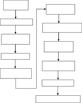
The segmentation of lung parenchyma is very challenging problem because homogeneity is not present in the lung region, similar densities in the pulmonary structures, different scanners and scanning protocols [20] and segmentation of lung regions will do the early diagnosis of lung cancer. The block diagram of the proposed method is shown in the below figure 1.
Read DICOM
contour model (SNAKES) to segment lung regions which is
used to locate the border of the object. One of the limitations of
SNAKE model is the construction of initial contour requires
often requires human interaction [7] and also iterations to be
specified to segment the desired object. Yang Liu et al [12]
have used thresholding and region growing algorithm to
segment lung regions. Nihad Mesanovic et al [13] have
suggested region growing as the algorithm to segment lung
regions. The limitation of region growing is its computational intensity and also performance is dependent on its initial seeds [7].
Zhaoxue Chen et al [6] used image threshold, fast flood filling technique, erosion operation and an area-filtering step to segment lung regions. Omid Talakoub et al [7] proposed a method which is based on detected lung edges by wavelet analysis, their proposed method is not only computational inexpensive but also robust and accurate in detecting lung boundaries. M.Arfan Jaffar et al [8] have proposed Genetic Algorithm (GA) and morphological operations to segment lung regions and the proposed method is completely an automated approach. Pei xiaomin Guo Hongyu Dai Jianping et al [11] have used region growing, thresholding and boundary correction algorithm based on convex hull and ray casting to extract lung parenchyma and the proposed method segments juxtapleural nodules (the nodule which is attached to lung wall) and pulmonary vessels by minimizing over- segmentation of adjacent regions such as the abdomen and
image
Apply thresholding
Label connected components
Clear borders
Apply Closure
Operation
Find the largest two areas
Apply Closure operation and fill image holes
Superimpose with original image
Smooth the borders of the image
Increase image size
Segmentation of lung regions
mediastinum.
Helen R et al [14] has proposed 2D Otsu algorithm based on
particle swarm optimization(PSO) is used to segment lung
parenchyma and the computation time of 2D Otsu is
approximately 90 seconds/slice and for proposed PSO
method is less than 1 second/slice . Thapaliya Kiran et al [15] used bit-plane technique, labelling, calculation of area for each label, boundary tracing algorithm to get lung regions. Diao Liming et al [16] have applied optimal threshold value,
boundary tracking and mathematical morphology method to segment lung parenchyma. Sivakumar S et al [17] proposed an algorithm called weighted Fuzzy C-Means (WFCM) to
Figure 1 Block diagram for the segmentation of lung regions
The overall process of the proposed method is as follows: Given an image, extract lung parenchyma by using a threshold of -420 HU [9]. Apply closure operation with the disk structuring element of radius 2 to close smaller area which is at the border of the lung. Find the connected components of all the areas in the image and then extract the two largest areas of the connected component.
Apply the closure operation with the disk structuring element of radius 12 [9] to close larger areas and fill the holes
IJSER © 2013 http://www.ijser.org
The research paper published by IJSER journal is about Automated Segmentation of Lung Regions using Morphological Operators in CT scan 1116
ISSN 2229-5518
inside the two connected components. Superimpose the resultant image with the given image to extract lung regions.
Many objects got extracted along with lung wall of the segmented lung regions and lung wall is removed by subtracting the eroded image from the image of step 7 from the algorithm [9]. Image erosion is done with the structuring element of shape disk which has a radius 2. Small lung nodules are not visible properly, so increase the image to visualize small lung nodules. Block diagram of the proposed method is shown in the figure 1.
The algorithm for the segmentation of lung region is as follows:
Step1: Read DICOM CT lung image.
Step2: Apply thresholding to segment lung parenchyma. Experimentally, threshold value was found at -420HU [9]
binary(i, j) = 1, if f(i, j) < 𝑇 ... (1)
0, otherwise
Here T is -420 HU [9].
binary(i,j) is the binary image after applying thresholding.
To convert Hounsfield Unit to gray level value uses the equation (2).
Gray Level value= 1024+ T ... (2)
Step 3: Label various connected components. R are the regions with different labels.
Step 4: Clear image border to remove the regions which have connected to the border of the image [5]
0, border(R) = B pixels
1, border(R) ≠ B pixels ... (3)
R are the regions with different labels. B pixels are the border
pixels of the image.
Step 5: Apply closure operation with disk which is of radius 2
𝐴 ●𝐵 = (𝐴⨁𝐵) ⊝ 𝐵 ... (4)
A is the input image and B is the structuring element.
Step 6: Find the two largest areas [5]
max�area(R)� lung1
12[9] and fill image holes.
Use the equation (4) to apply closure operation
Step 8: Super impose the image of step 7 with input image and the segmented image is as shown in the figure 3.
Step 9: Smoothen the borders of the image [9] by eroding the image from step 7 with the structuring element of shape disk which has a radius of 2 and subtracting an eroded image from the image of step 7.
𝐴ϴ𝐵 = {𝑧|(𝐵)𝑧 ⊆ A} ... (6)
A is the reference binary image and B is the structuring
element of shape disk with the size 2.z is the outcome element
of the equation (6).
S=A-z ... (7) S contains image which has smooth borders. Step 10: Increase the image size.
4 RESULTS
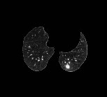
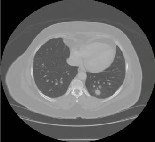
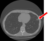
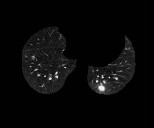
Nihad Mrsanovic et al [13] method applied on figure 2(a) and
2(b). In figure 2(a), the upper image is the input image and
lower image is the segmentation of lung regions which is not
having lung nodules attached to the lung wall. In figure2(b),
lung nodule is attached to the lung wall where upper image is
the input image and lower image is the segmentation of lung
regions which has an arrow mark to locate lung nodule is
missing in the segmentation of the lung regions.
The limitation of Nihad Mrsanovic et al [13] method is that it
is not segmenting the lung nodule which is shown in the
figure 2(b).Segmented image of 2(a) and 2(b) has the borders
with lung wall and image size is not increased.
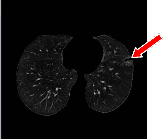
To overcome the problem in figure 2(b), thresholding and morphological operators have used which is applied on figure
3(a) and 3(b). In figure 3(a) upper image and lower image are the input image and the segmented image which has no lung nodules at the lung borders.
In figure 3(b), lung nodule is attached at left lung of the upper image and lower image which is shown with arrow mark. In figure 3(b), lung regions are segmented along with the lung nodule attached to the border of the left lung. Proposed method is applied to smooth the borders of segmented image and to increase the image size.
5 CONCLUSION
Lungregion = �
(max −1)�area(R)� lung2
... (5)
Nihad Mrsanovic et al [13] used region growing technique which cannot segment the nodules attached to the borders of
The first largest connected component is lung1 and second
largest connected component is lung2. The connected
components which are less than lung2 has been removed by
using equation (5).
Step 7: Apply closure operation with disk which is of radius
the lung image. The proposed method is an automated approach which segments the lung regions along with nodules which are attached to lung borders of the image using thresholding and morphological operators to assist radiologist for the better diagnosis of lung cancer.
IJSER © 2013 http://www.ijser.org
The research paper published by IJSER journal is about Automated Segmentation of Lung Regions using Morphological Operators in CT scan 1117
ISSN 2229-5518
The Proposed method removes the lung walls of the input image and increases the size of the image to visualize small lung nodules.

(a) (b)
Figure 2 The upper, lower images are input and segmented
images with different two cases in which lung nodule is not attached to the lung wall (a) and the lung nodule is attached to the lung wall (b). Results are from Nihad Mrsanovic’s [13] method.
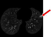
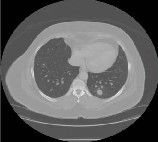
(a) (b)
Figure 3 The upper, lower images are input and segmented images with different two case in which lung nodule is not attached to the lung wall (a) and the lung nodule is attached to the lung wall (b). Results are from the proposed method
REFERENCES
[1] Austin J H M ,Mueller N L, Friedman P J, Hansell D M, Naidich D P, Remy-Jardin M, Webb W R, Zerhouni E A, ”Glossary of terms for CT of the lungs: recommendation of the nomenclature committee of the fleischner society”, Radiology 1996,200 pp.327-331.
[2] Shiying Hu, Eric A. Hoffman, and joseph M.Reinhardt,” Automatic lung segmentation for accuracte quantitation of volumetric x-ray ct images”,IEEE Trans.Med.Imaging,Vol.20,no.6,pp.490-498,June 2001.
[3] G. Samuel, III.Armato, M. Geoffrey ,F.Michael, R.Charles, Y.David,
”Lung image database consortium-developing a resource for the medical imaging research community”,Radiology 24(2004) 739-748.
[4] Yoshinori Itai, Hyoungseop Kim, Seiji Ishikawa,Shigehiko Katsura gawa, Takayuki, Katsumi Nakamura and Akiyoshi Yamamoto,
”Automatic Segmentation of Lung Areas Based on SNAKES and Extraction of Abnormal Area”, Proceedings of the 17th IEEE International Conference on Tools with Artificial Intelligence(ICTAI’05),2005.
[5] Aamir Saeed Malik,Tae-Sun Choi,”A novel algorithm for segmentation of lung images”,7th International symposium,IS BMDA
2006,Thessaloniki,Greece,December 7-8,2006,proceedings.
[6] Zhaoxue Chen,Xiwen Sun,Shengdong Nie, ”An Efficient Method of Automatic Pulmonary Parenchyma Segmentation in CT Images”, Proceedings of the 29th Annual International Conference of the IEEE EMBS, Cite Internationale, Lyon, France August 23-26,2007.
[7] Omid Talakoub,Javad Alirezaie,Paul Babyn,”Lung Segmentation in
Pulmonary CT Images Using Wavelet Transform”,ICASSP,IEEE,2007 [8] M. Arfan Jaffar et al,”GA and morphology based automated
segmentation of lungs from CT scan images”,IEEE,2008.
[9] Eng. Michael Samir Labib Habib,”A computer aided diagnosis system (CAD) for the detection of pulmonary nodules on CT scans”,systems and biomedical engineering department,faculty of engineering,cairo university,Giza,Egypt,2009.
[10] Lin-Yu Tseng,Li-Chin Huang,”An Adaptive Thresholding Method for Automatic Lung Segmentation in CT Images”IEEE AFRICON
2009.
[11] Pei xiaomin Guo Hongyu Dai Jianping, Dai Jianping,”A Novel Lung Segmentation and Boundary Correction Method”,2nd International Conference on Biomedical Engineering and Informatics, BMEI, IEEE,
2009
[12] Yang Liu,Zhian Xing,Chao Deng,Ping Li and Maozu Guo,”Automatically Detecting Lung Nodules Based on Shape Descriptor and Semi-Supervised Learning”,International Conference on Computer Application and System Modeling (ICCASM ),IEEE,
2010.
[13] Nihad Mesanovic,Mislav Grgic,Haris Huseinagic,Matija Males,Emir Skejic,Muamer Smajlovic,”Automatic segmentation of the lungs with region growing algorithm”,IWSSIP,June 2011
[14] R.Helen,Dr.N Kamaraj,Dr.K.Selvi,V.Raja Raman,”Segmentation of Pulmonary Parenchyma in CT Lung Images based on 2D Otsu optimized by PSO”,Proceedings of ICETECT ,IEEE.2011.
[15] Thapaliya Kiran and Goo-Rak Kwon,”An Advanced Segmentation Using Bit-Plane Slicing Technique in Extraction of Lungs Region”,Second Asian Himalayas International Conference on Internet(AH-ICI)IEEE,2011.
[16] Diao Liming,Zhang Caiming,Gao Shanshan,”A New Algorithm of Automatic Lung Parenchyma Segmentation Based on CT Images”, International Conference on Consumer Electronics,Communications and Networks(CECNet), IEEE,2011.
IJSER © 2013 http://www.ijser.org
The research paper published by IJSER journal is about Automated Segmentation of Lung Regions using Morphological Operators in CT scan 1118
ISSN 2229-5518
[17] Sivakumar S and Chandrasekar C,”Lungs Image Segmentation through Weighted FCM”,International Conference on Recent Advances in Computing and Software Systems,IEEE,2012.
[18] American Cancer Society,Cancer Facts and Figures,American Cancer
Society,NewYork,NY,USA,2013.
[19] http://imaging.cancer.gov/programsandresources/informationsystem s/lidc
[20] Ayman El-Baz,Garth M.Beache,Georgy Gimel farb,Kenji Suzuki,Kazunori Okada,Ahmed Elnakib,Ahmed Soliman and Behnoush Abdollahi,”Computer-Aided Diagnosis Systems for Lung Cancer; Challenges and Methodologies”Review Article, International Journal of Biomedical Imaging,Volume 2013,Article ID 942353,46 pages.
IJSER © 2013 http://www.ijser.org